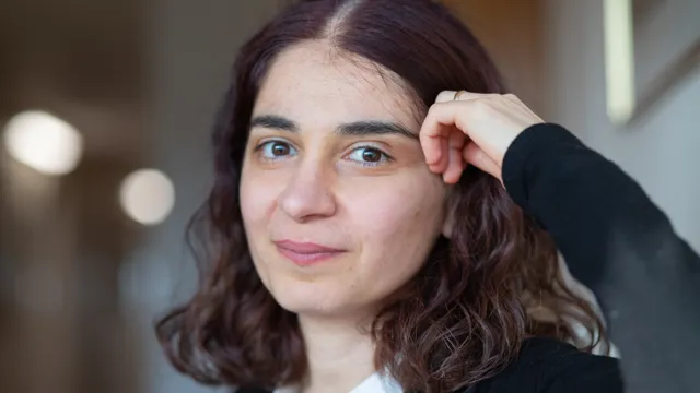
During her research education at the Department of Clinical and Experimental Medicine, Kjersti Tunströmer has improved the methods researchers use to investigate platelets. Platelets, also known as “thrombocytes”, are a type of blood cell whose most important function is to plug minor damage to the blood vessel and in this way prevent bleeding and a shortage of oxygen. Platelets, however, can sometimes cause problems.
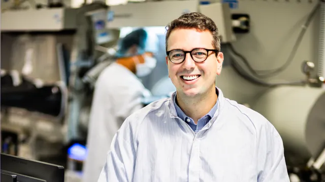
“Platelets play a central role in the formation of blood clots, which in turn are the most common cause of myocardial infarction and stroke. Drugs to inhibit platelet function are a very important means of preventing and treating these diseases”, says Professor Tomas Lindahl.
Artificial “wounds”
The research group uses narrow channels, known as “flow chambers”, that mimic blood vessels. An artificial “wound” is created in the flow chamber, or – to be more accurate – a point is created with the same proteins that come into contact with the blood when a blood vessel is damaged. The researchers pump blood through the flow chamber, and when the platelets encounter the “damage”, they form clumps and a tiny plug that seals the hole in the blood vessel. By staining the platelets with fluorescent dyes, the researchers can study the process in a microscope and record huge numbers of images, containing enormous amounts of information.The formation of a blood clot is studied in microscope. Photo credit Karin Söderlund Leifler
Flow chambers in themselves are nothing new, and are used by many research groups around the world. In the work described in her thesis, Kjersti Tunströmer has developed a new method and a computer program to interpret the microscope images from experiments in flow chambers. The information from the images can be used to create 3-dimensional images and video films of the clot, or images that correspond to a cross-section through it.
“Our method allows us to follow thousands of platelets as they move during the process in which blood clots form. An important process in the formation of a blood clot is the way in which the platelets approach each other. And using the method, we can study how different drugs influence platelet motion”, says Kjersti Tunströmer.
The method is described in an article that has been published in the journal Thrombosis and Haemostasis, where videos created by the researchers have been published.
The article: "", Tunströmer K, Faxälv L, Boknäs N et al., Thromb Haemost 2018; 118(09): 1600-1611, doi: 10.1055/s-0038-1668151
Translation by George Farrants